
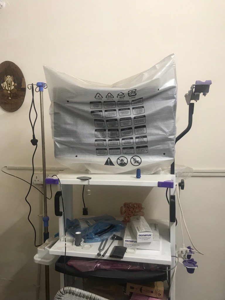
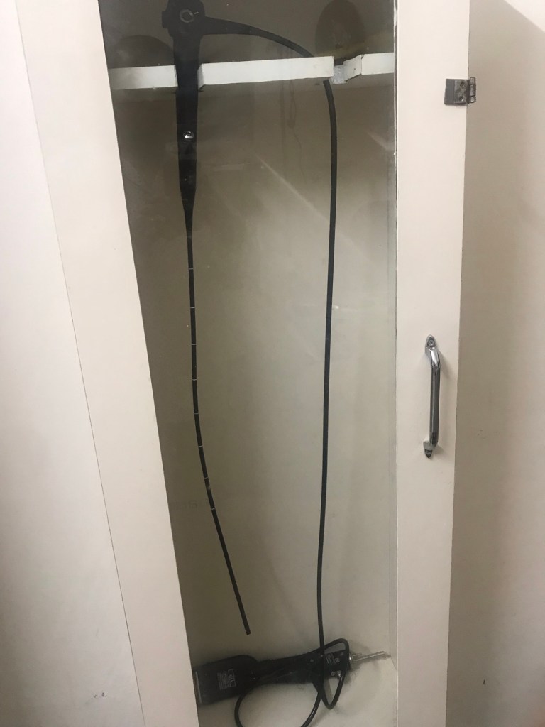
Display of fibrooptic Brochoscope
We are routinely doing fibroptic Brochoscopy both diagnostic and therapeutic on Olympus fibroptic Broncoscope.
A fiber optic bronchoscope is a critical tool in diagnosing and managing chest diseases, allowing direct visualization of the airways and lungs. Below is a concise overview of its role in chest disease, tailored to your query:
Role in Chest Diseases
- Diagnostic Applications:
- Lung Cancer: Visualizes tumors, obtains biopsies (forceps, needle aspiration), or performs bronchial washings for cytology. Fluorescent bronchoscopy detects early-stage lesions.
- Infections: Identifies causes of pneumonia, tuberculosis, or fungal infections via bronchoalveolar lavage (BAL) or protected brush samples.
- Interstitial Lung Diseases: BAL and transbronchial biopsies help diagnose sarcoidosis, pulmonary fibrosis, or hypersensitivity pneumonitis.
- Hemoptysis: Locates bleeding sources (e.g., tumors, bronchiectasis) for targeted intervention.
- Airway Abnormalities: Assesses tracheal stenosis, bronchomalacia, or foreign body presence.
- Mediastinal Disorders: Endobronchial ultrasound (EBUS)-guided bronchoscopy samples lymph nodes for staging lung cancer or diagnosing lymphoma.
- Therapeutic Applications:
- Foreign Body Removal: Extracts aspirated objects using forceps or baskets.
- Airway Obstruction: Clears mucus plugs or blood clots in conditions like COPD or atelectasis.
- Bleeding Control: Instills epinephrine or uses balloon tamponade for hemoptysis.
- Stent Placement: Deploys stents for airway narrowing due to tumors or scarring.
- Difficult Intubation: Guides endotracheal tube placement in patients with airway distortion (e.g., from tumors or trauma).
- Laser Therapy: Delivers laser via bronchoscope to debulk tumors in advanced lung cancer.
Advantages in Chest Disease
- Minimally Invasive: Performed under local anesthesia with sedation, reducing risks compared to surgical alternatives.
- Real-Time Visualization: Flexible tip (180° up, 130° down angulation) navigates complex bronchial anatomy.
- Versatility: Combines diagnostic (biopsy, BAL) and therapeutic (stenting, foreign body removal) capabilities in one procedure.
- Outpatient Setting: Often done bedside or in endoscopy suites, minimizing hospital stays.
Specific Chest Conditions Addressed
- Chronic Obstructive Pulmonary Disease (COPD): Assesses airway inflammation, clears secretions, or guides bronchial thermoplasty.
- Bronchiectasis: Evaluates extent, collects samples to identify pathogens.
- Pulmonary Alveolar Proteinosis: Performs whole-lung lavage to remove surfactant buildup.
- Asthma: Investigates severe cases or guides bronchial thermoplasty for refractory asthma.
Procedure Details
- Setup: Flexible fiber optic bronchoscope (5.0-6.0 mm for adults, smaller for pediatrics) with light source (LED/halogen) and working channel (1.2-3.2 mm for suction/biopsy).
- Anesthesia: Lidocaine (spray or liquid) for airway numbing; sedation (midazolam, fentanyl) for comfort.
- Duration: 15-60 minutes, depending on complexity (e.g., biopsy vs. simple inspection).
- Risks: Rare but include bleeding, pneumothorax (with biopsy), or transient hypoxia.
Limitations
- Image Quality: Inferior to videobronchoscopes (which use CCD/CMOS chips).
- Fragility: Fiber optic bundles can break, requiring costly repairs.
- Learning Curve: Requires skilled operators for complex interventions like EBUS or laser therapy.
- Thanks
If you need details on a specific chest disease or procedure, let me know!


")


